 Weather today
Google helps map one cubic millimeter of human brain tissue that comes in at 1.4 petabytes

Google, in collaboration with Harvard University, today released a browsable 1.4-petabyte reconstruction of a very small part of the human cortex.
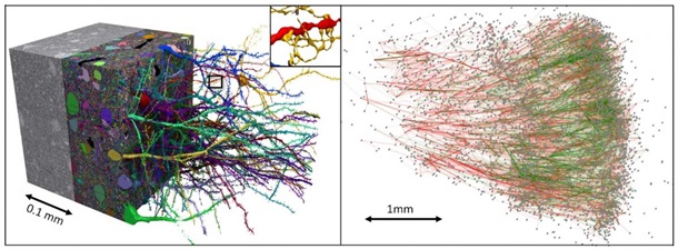
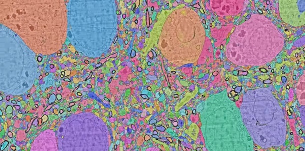
The dataset comprises imaging data that covers roughly one cubic millimeter of brain tissue, and includes tens of thousands of reconstructed neurons, millions of neuron fragments, 130 million annotated synapses, 104 proofread cells, and many additional subcellular annotations and structures, 9to5google.com reports.
The cerebral cortex is a thin surface layer of the brain that plays a crucial role in thinking, memory, planning, perception, language, attention, and most other higher-level cognitive functions.
Although there has been some progress in understanding the macroscopic organization of this very complicated tissue, its organization at the level of individual nerve cells and their interconnecting synapses is largely unknown.
This sample tissue was anonymously donated from patients that have undergone surgery to treat epilepsy at the Massachusetts General Hospital in Boston (MGH). It was then given to researchers at Harvard’s Lichtman laboratory.
The Harvard researchers cut the tissue into ~5300 individual 30 nanometer sections using an automated tape collecting ultra-microtome, mounted those sections onto silicon wafers, and then imaged the brain tissue at 4 nm resolution in a customized 61-beam parallelized scanning electron microscope for rapid image acquisition.
The end result was 225 million individual 2D images that Google then computationally stitched and aligned into a 3D volume with thousands of Google Cloud TPUs were leveraged in the process. This human brain map is now accessible through Google’s web-based Neuroglancer visualization tool.








 Studio One